Vimentin Antibody

Cat# AX10005

Size : 400ul

Brand : Abcepta

Request more information

Contact local distributor :


Phone : +1 850 650 7790

  • IF - Vimentin Antibody AX10005
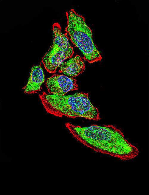
    Confocal immunofluorescent analysis of Vimentin Antibody with A549 cell followed by Alexa Fluor 488-conjugated goat anti-rabbit lgG (green). Actin filaments have been labeled with Alexa Fluor 555 phalloidin (red). DAPI was used to stain the cell nuclear (blue).
  • IF - Vimentin Antibody AX10005
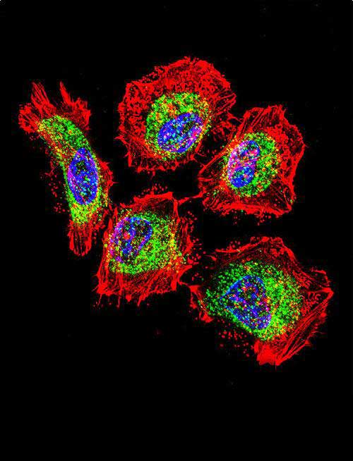
    Confocal immunofluorescent analysis of Vimentin Antibody with Hela cell followed by Alexa Fluor 488-conjugated goat anti-rabbit lgG (green). Actin filaments have been labeled with Alexa Fluor 555 phalloidin (red). DAPI was used to stain the cell nuclear (blue).
  • IF - Vimentin Antibody AX10005
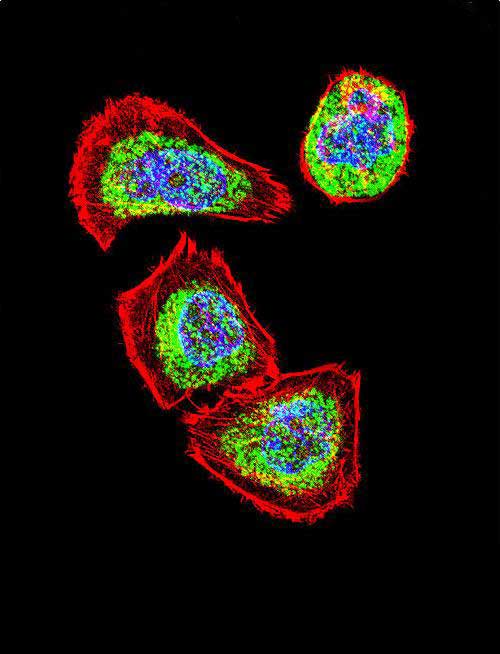
    Confocal immunofluorescent analysis of Vimentin Antibody with U251 cell followed by Alexa Fluor 488-conjugated goat anti-rabbit lgG (green). Actin filaments have been labeled with Alexa Fluor 555 phalloidin (red). DAPI was used to stain the cell nuclear (blue).
  • WB - Vimentin Antibody AX10005
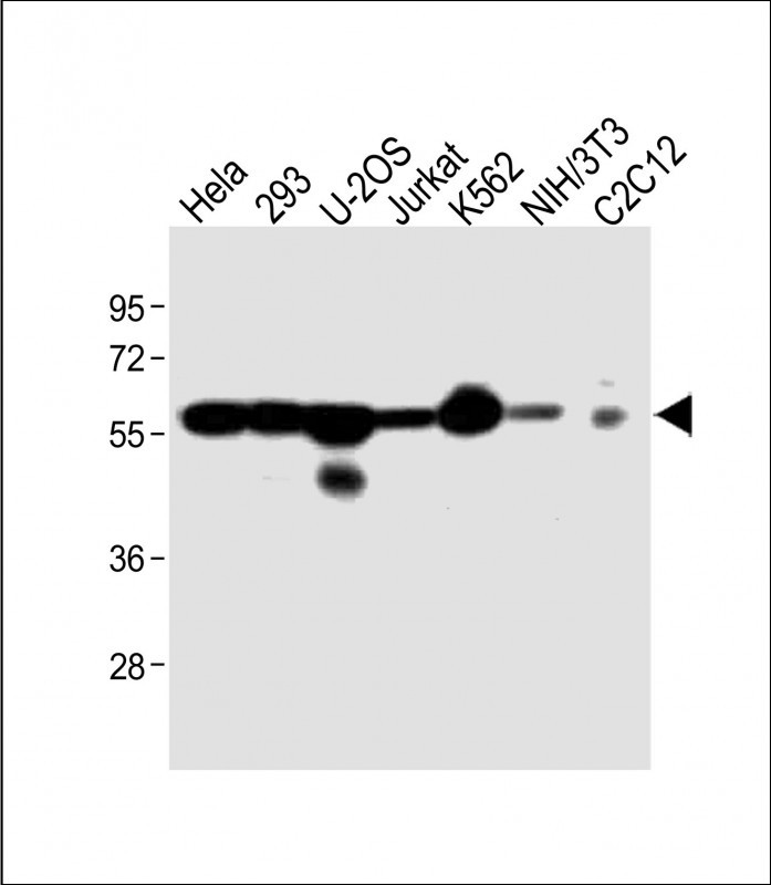
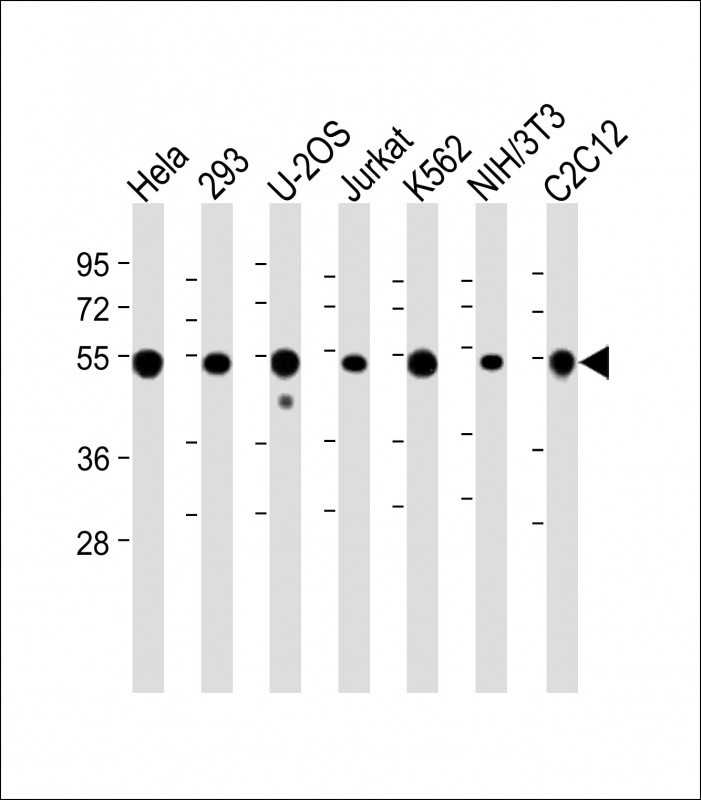
    All lanes : Anti-VIME Antibody at 1:4000 dilution Lane 1: Hela whole cell lysate Lane 2: 293 whole cell lysate Lane 3: U-2OS whole cell lysate Lane 4: Jurkat whole cell lysate Lane 5: K562 whole cell lysate Lane 6: NIH/3T3 whole cell lysate Lane 7: C2C12 whole cell lysate Lysates/proteins at 20 µg per lane. Secondary Goat Anti-Rabbit IgG, (H+L), Peroxidase conjugated at 1/10000 dilution. Predicted band size : 54 kDa Blocking/Dilution buffer: 5% NFDM/TBST.
  • WB - Vimentin Antibody AX10005
    All lanes : Anti-VIME Antibody at 1:2000 dilution Lane 1: Hela whole cell lysate Lane 2: 293 whole cell lysate Lane 3: U-2OS whole cell lysate Lane 4: Jurkat whole cell lysate Lane 5: K562 whole cell lysate Lane 6: NIH/3T3 whole cell lysate Lane 7: C2C12 whole cell lysate Lysates/proteins at 20 µg per lane. Secondary Goat Anti-Rabbit IgG, (H+L), Peroxidase conjugated at 1/10000 dilution. Predicted band size : 54 kDa Blocking/Dilution buffer: 5% NFDM/TBST.
  • IHC-P-Leica - Vimentin Antibody AX10005
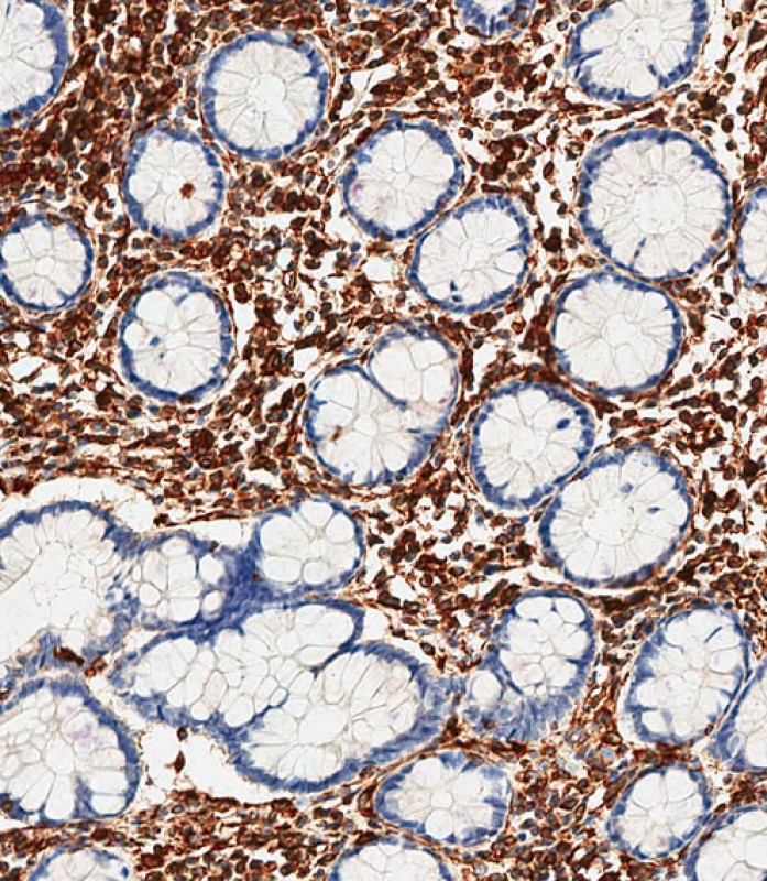
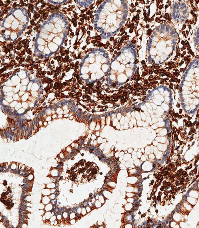
    Immunohistochemical analysis of paraffin-embedded Human small intestine tissue using AX10005 performed on the Leica® BOND RXm. Tissue was fixed with formaldehyde at room temperature, antigen retrieval was by heat mediation with a EDTA buffer (pH9. 0). Samples were incubated with primary antibody(1:500) for 1 hours at room temperature. A undiluted biotinylated CRF Anti-Polyvalent HRP Polymer antibody was used as the secondary antibody.
  • IHC-P-Leica - Vimentin Antibody AX10005
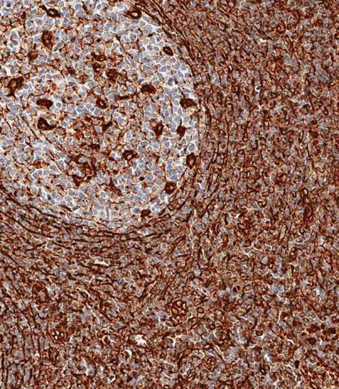
    Immunohistochemical analysis of paraffin-embedded Human tonsil tissue using AX10005 performed on the Leica® BOND RXm. Tissue was fixed with formaldehyde at room temperature, antigen retrieval was by heat mediation with a EDTA buffer (pH9. 0). Samples were incubated with primary antibody(1:500) for 1 hours at room temperature. A undiluted biotinylated CRF Anti-Polyvalent HRP Polymer antibody was used as the secondary antibody.
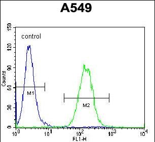
  • FC - Vimentin Antibody AX10005
    Vimentin Antibody flow cytometric analysis of A549 cells (right histogram) compared to a negative control cell (left histogram).Alexa Fluor 488-conjugated donkey anti-rabbit lgG secondary antibodies were used for the analysis.
Product Information
Application
  • Applications Legend:
  • WB=Western Blot
  • IHC=Immunohistochemistry
  • IHC-P=Immunohistochemistry (Paraffin-embedded Sections)
  • IHC-F=Immunohistochemistry (Frozen Sections)
  • IF=Immunofluorescence
  • FC=Flow Cytopmetry
  • IC=Immunochemistry
  • ICC=Immunocytochemistry
  • E=ELISA
  • IP=Immunoprecipitation
  • DB=Dot Blot
  • CHIP=Chromatin Immunoprecipitation
  • FA=Fluorescence Assay
  • IEM=Immuno electron microscopy
  • EIA=Enzyme Immunoassay
WB, IF, FC, IHC-P-Leica, E
Primary Accession P08670
Other Accession NP_003371.2
Reactivity Human, Mouse
Host Rabbit
Clonality Polyclonal
Isotype Rabbit IgG
Clone/Animal Names RB41034
Calculated MW 53652 Da
Additional Information
Gene ID 7431
Other Names Vimentin, VIM
Target/Specificity This Vimentin antibody is generated from rabbits immunized with human Vimentin recombinant protein.
Dilution IF~~1:50
WB~~1:2000
IHC-P-Leica~~1:500
FC~~1:50
Format Purified polyclonal antibody supplied in PBS with 0.09% (W/V) sodium azide. This antibody is purified through a protein A column, followed by peptide affinity purification.
StorageMaintain refrigerated at 2-8°C for up to 2 weeks. For long term storage store at -20°C in small aliquots to prevent freeze-thaw cycles.
PrecautionsVimentin Antibody is for research use only and not for use in diagnostic or therapeutic procedures.
Protein Information
Name VIM (HGNC:12692)
Function Vimentins are class-III intermediate filaments found in various non-epithelial cells, especially mesenchymal cells. Vimentin is attached to the nucleus, endoplasmic reticulum, and mitochondria, either laterally or terminally. Plays a role in cell directional movement, orientation, cell sheet organization and Golgi complex polarization at the cell migration front (By similarity). Protects SCRIB from proteasomal degradation and facilitates its localization to intermediate filaments in a cell contact-mediated manner (By similarity).
Cellular Location Cytoplasm. Cytoplasm, cytoskeleton. Nucleus matrix {ECO:0000250|UniProtKB:P31000}. Cell membrane {ECO:0000250|UniProtKB:P20152}
Tissue Location Highly expressed in fibroblasts, some expression in T- and B-lymphocytes, and little or no expression in Burkitt's lymphoma cell lines. Expressed in many hormone-independent mammary carcinoma cell lines.
Research Areas
Citations ( 0 )

Background

This gene encodes a member of the intermediate filament family. Intermediate filamentents, along with microtubules and actin microfilaments, make up the cytoskeleton. The protein encoded by this gene is responsible for maintaining cell shape, integrity of the cytoplasm, and stabilizing cytoskeletal interactions. It is also involved in the immune response, and controls the transport of low-density lipoprotein (LDL)-derived cholesterol from a lysosome to the site of esterification. It functions as an organizer of a number of critical proteins involved in attachment, migration, and cell signaling. Mutations in this gene causes a dominant, pulverulent cataract.

References

Kers, J., et al. Transplantation 90(5):502-509(2010)
Pinheiro, A.P., et al. Am. J. Med. Genet. B Neuropsychiatr. Genet. 153B (5), 1070-1080 (2010) :
Korita, P.V., et al. Anticancer Res. 30(6):2279-2285(2010)
Martins-de-Souza, D., et al. J Psychiatr Res (2010) In press :
Li, M., et al. J. Exp. Clin. Cancer Res. 29, 109 (2010) :

Availability: In stock
Bulk Size
Bulk Order
Call
Call
Cat# AX10005

You might also be interested by the following products:


Cat#
Description
Cond.
Price Bef. VAT