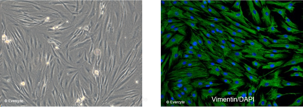
Evercyte’s human dermal fibroblast cell line fHDF/TERT166 can be grown without limitations while maintaining expression of cell type specific markers and functions. Therefore, these cells are frequently used as standardized in vitro model to study processes involving fibroblasts such as formation of the extracellular matrix, inflammation, wound healing or fibrosis. Moreover, the cells embedded into a collagen matrix and co-cultured with telomerized keratinocytes allow the establishment of standardizable 3D skin equivalents.
General information
Cat#: CHT-031-0166
Morphology and marker expression

fHDF/TERT166 cells are characterized by the typical, spindle shaped morphology of mesenchymal cells and homogenously express the fibroblast marker Vimentin. Cell nuclei are counterstained with DAPI.
Response to cytokines / expression of IL-6 upon IL-17A treatment

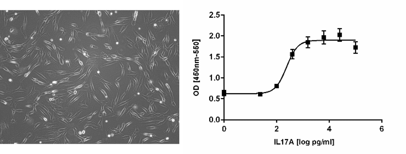
Treatment of telomerized human dermal fibroblasts fHDF/TERT166 with Interleukin-17A (IL17A) results in expression of interleukin-6 (IL6) in a concentration dependent manner.
Myofibroblast differentiation / effect of extracellular vesicles

Treatment of telomerized human dermal fibroblasts fHDF/TERT166 with transforming growth factor beta (TGF-ß) induces expression of alpha smooth muscle actin (𝛂-SMA). Treatment of cells with TGF-ß together with extracellular vesicles (EVs) from mesenchymal stem cells reduces 𝛂-SMA expression significantly, demonstrating an effect of EVs on myofibroblast differentiation.
Cell migration / induction of fibroblast growth upon treatment with extracellular vesicles

fHDF/TERT166 were seeded into chamber slides and a physical gab within the monolayer was created followed by monitored the process of cell migration into the gap. Significantly less free area between the cells was detected upon addition of extracellular vesicles from mesenchymal stem cells.


Follow us on Social Media